Distal Femoral Osteotomy Seattle, Wa
All authors offered crucial feedback and helped form the analysis, evaluation and manuscript. The anonymised results of the radiological measurements and the medical questionnaires are connected within the type of an Excel spreadsheet. The common deviation of the ultimate HKA compared to the preoperative planning was 2.4° ± 0.four°. On discharge from hospital you will have a 2 week course of clexane injections to thin the blood and scale back the risk of a DVT whilst you’re less cellular than ordinary.
With the affected person positioned in the supine position on a radiolucent desk, the articular surface was recognized by palpation and radioscopy. An incision of ∼ 10 cm was performed, extending proximally from the medial knee joint line. Next, the vastus medialis was bluntly dissected to expose the condyle and the medial femoral cortex. Thus, no neurovascular structure was uncovered or put at risk through the surgical entry, and the bone floor required for osteotomy was safely approached.
Smoking has a profound impact on fracture healing and we should not risk the bone not therapeutic back collectively. Patients who are overweight usually find their knee pain is considerably improved when they shed weight. Simple analgesia similar to paracetamol along with ibuprofen may help with pain and sleep disturbance type the ache. Limb realignment can even assist relieve pain and problems arising from a patella that’s not gliding usually across the tip of the femur. This is an operation normally carried out for arthritis and occasionally patella instability problems across the knee.
This article provides a detailed, step-wise methodology that permits the reproducible creation of a medial closing-wedge DFO for the valgus knee utilizing locking-plate fixation. Both medial closing-wedge and lateral opening-wedge osteotomies of the distal femur have been reported for correction of genu valgum.5 Patient-reported knee quality of life is improved by both method.6, 7, 8, 9 Advantages of every method are detailed in Table 1. The incidence of femoral distal development plate fractures is considered to be roughly 1 to six% of all growth plate fractures .
Our Osteotomy Plates
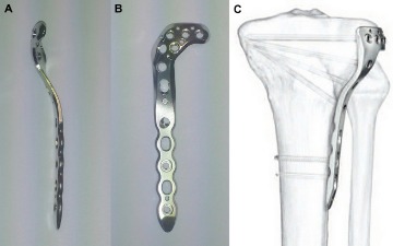
If performing a bigger correction, it’s useful to perforate the medial cortex with a drill bit to allow a controlled opening. Corticocancellous wedges are harvested from the femoral neck portion of an allograft femoral head and positioned into the osteotomy web site according to the preoperative plan. These wedges stabilize the osteotomy while the ultimate mechanical axis views are verified with fluoroscopy . The distal, lateral femoral locking plate is then positioned on the lateral femoral cortex.

A wedge-formed bone graft is removed from the pelvic bone and inserted to fill the osteotomy defect or donated cadaver bone is used. Once the right alignment of your leg is confirmed, the muscles and blood vessels are launched and the incision is sutured. Intraoperative alignment control was carried out with the x-ray grid, a 3 mm skinny phenolic resin hard paper plate with intersected distinguishable radiopaque reference lines for determination of the mechanical axis. At the start of the procedure, meniscal and cartilage lesions had been evaluated with arthroscopy. Only TomoFix plates have been used as implants for the oHTO and the operative method was much like Staubli et al. with biplanar cutting approach .
What’s Distal Femoral Osteotomy?
This can occur after an damage to the crescent moon formed shock absorbers in the knee often known as menisci, significantly the lateral meniscus. An injury to the ACL could make the knee less stable and prone to cartilage injury over time. An incision is made over the distal femur the place the osteotomy is to be carried out.
A diagnostic arthroscopy can be carried out to verify that there’s isolated lateral compartment illness . Concomitant procedures could be performed right now to handle lateral compartment chondral or meniscal disease or deficiency. Two surgical method choices could be thought of for a lateral, distal femoral osteotomy. The first is a true further-articular strategy by which a 12- to 15-cm lateral incision is revamped the midline lateral femur and angulated anterior 2 cm distal to lateral epicondyle. The vastus lateralis is elevated from intermuscular septum, being cautious to coagulate arterial branches of the profunda femoris. Very promising outcomes have recently been published by a single research group utilizing patient-specific slicing guides in oHTO and oDFO .
- The wedge guidewire was positioned with the angular reduce predefined for every case, and ∼ 75% of the wedge was sectioned and removed; this was thought of a partial procedure.
- In the immediate postoperative interval, all sufferers are placed on a chemical deep vein thrombosis prophylaxis agent, based mostly on preoperative risk components.
- In this circumstance, a more anterior skin incision, adopted by a formal arthrotomy, was carried out, as a concomitant lateral femoral condyle osteochondral allograft switch was performed.
- These wires additionally serve as a boundary to information the saw blade and make sure that over-resection doesn’t occur.
- This is defined by the technically demanding closed wedge osteotomy, because the surgeon must depend on the accuracy of the bone resection, and intraoperative readjustment is just possible to a limited extent .
There have been no relevant differences in hospital stay, blood loss or surgery time. One occurrence of delayed bone formation in the oHTO group was efficiently treated with autologous bone grafting. On average, ultimate radiological examination took place 6 months after implant removal, together with LSR and lateral x-ray, which was typically 18 months postoperative. Mean observe up for clinical examination including questionnaires (Lysholm score, SF-36, VAS) was forty seven months postoperatively (Tab. 2), with a minimal of 24 months. Patients will proceed to see enchancment in the knee symptoms over the 12 months after their operation. Our patient database suggests that the majority affected person’s symptoms proceed to improve slowly long after that as nicely.
Most patients have their operation accomplished underneath a spinal anaesthetic with some sedation. This includes an injection within the back to numb the legs which supplies ache aid even after the operation has completed. Usually we’ll begin the procedure by performing an arthroscopy of the knee joint.